NeuroNavigator
| Category | Price | Seller | Device |
|---|---|---|---|
| Education | Free | imeka app | iPhone, iPad, iPod |


Visualize and interact with human brain white matter, gray matter, and vascular anatomy derived from cutting edge multi-modal human neuroimaging magnetic resonance imaging (MRI) datasets.
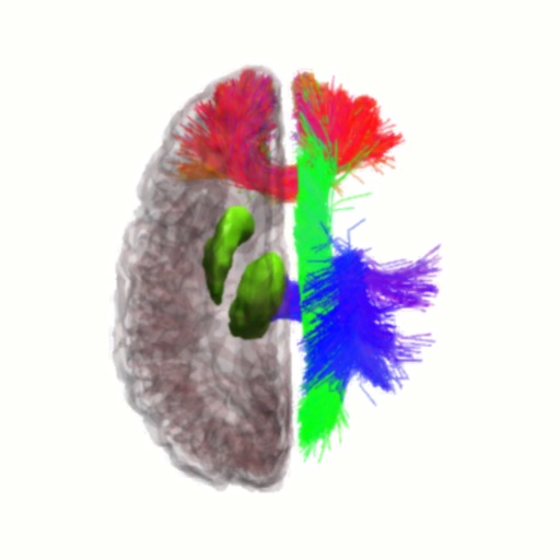
The app contains neuroimaging data from a 33 year old healthy human male brain including:
- 84 cortical areas based on gray matter segmentation of anatomical T1-weighted MRI image (T1)
- 24 subcortical areas, including ventricles and cerebellum segmented from anatomical T1
- 33 white matter pathways reconstructed using tractography from diffusion weighted MRI image (DWI)
- venous segmentation based on susceptibility weighted MRI image (SWI)
- arterial segmentation based on time of flight MRI image (TOF)
- functional connectivity based on blood oxygen level dependent (BOLD) functional MRI images
Cortical and subcortical regions of interest (ROIs) were segmented automatically using the Freesurfer software package:
https://www.sciencedirect.com/science/article/pii/S1053811912000389
White matter pathways were processed and automatically segmented using in-house software developed at Imeka, based on the following publication:
https://www.sciencedirect.com/science/article/pii/S1053811917305839
Venous and arterial segmentations were segmented automatically using in-house software developed at the University of Sherbrooke, based on the following publication:
https://onlinelibrary.wiley.com/doi/abs/10.1002/hbm.24337
All data was acquired with informed consent on a 3 Tesla Philips Ingenia MRI scanner at the Centre Hospitalaire Universite de Sherbrooke in Sherbrooke, Quebec, Canada.
Useful for learning about the anatomy of the brain.